
Szczepanik Research Group
Department of Theoretical Chemistry
Faculty of Chemistry, Jagiellonian University
Gronostajowa 2, 30-387 Krakow, Poland
Tel: (+48) 12 686 23 90
E-mail: dariusz.szczepanik@uj.edu.pl
HOME PROJECTS PUBLICATIONS runEDDB X
About EDDB Download Tutorials Manual
◼ Tutorial 6 — π-Reorganization in a hydrogen-bonded guanine quartet
Four guanines, eight hydrogen bonds, one planar ring. Does hydrogen bonding change the π-system of each guanine, and where? The differential BDF map of Tutorial 5 answers it again — with a new twist: the reference is a single molecule, the complex holds four, and only one of them can be aligned at a time.
Before you start
A guanine quartet (G-quartet) is a planar, cyclic assembly of four guanines — the building block of G-quadruplex DNA. Each guanine donates two hydrogen bonds from its Watson–Crick edge (N1–H and N2–H) to one neighbour, and accepts two at its Hoogsteen edge (O6 and N7) from the other: eight hydrogen bonds in all, N1–H···O6 and N2–H···N7. The quartet studied here has four-fold (C4h) symmetry, so all four guanines are equivalent.
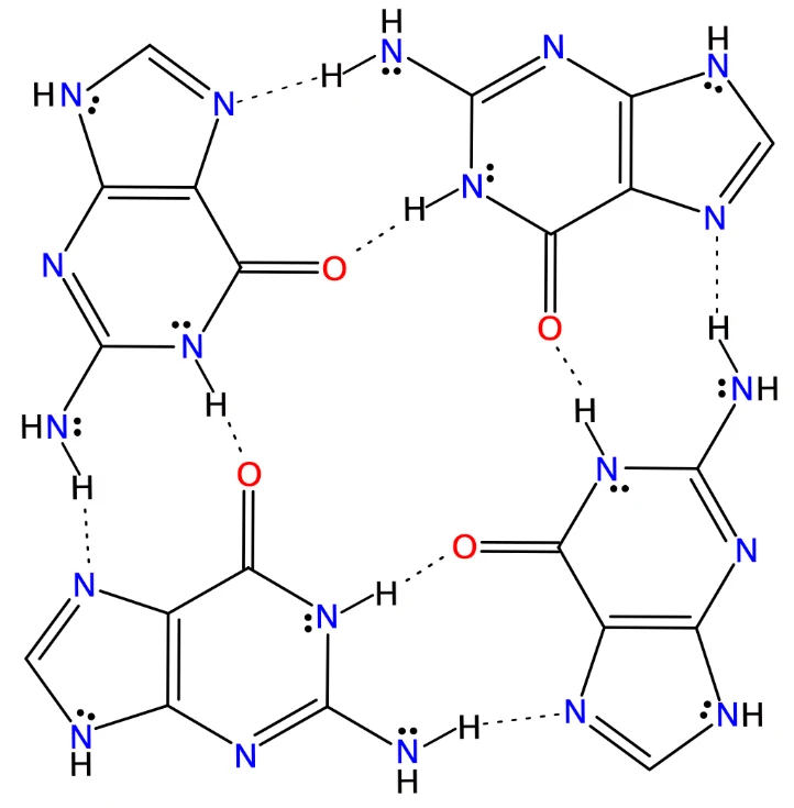
Figure 1. The G-quartet: four guanines held by eight N–H···O and N–H···N hydrogen bonds. The π-conjugated lone pairs are drawn as dots.
You need two geometry files: G.xyz, an isolated guanine, and G4.xyz, the quartet. In G4.xyz the four guanines are listed as consecutive blocks of 16 atoms, each in the same order as G.xyz.
Our aim is a differential map that shows how the π-structure of each guanine changes when the quartet forms:
ΔBDFπ = BDFπ(G4) − BDFπ(G)
Guanine has 14 π-electrons: eight from four double bonds and six from three π-conjugated lone pairs (Figure 1). The quartet has 56. Hence there are 7 π-NOBDs for the monomer and 28 for the quartet.
Part 1 — The reference: BDFπ of an isolated guanine
- -s 1:7 — dissect the seven π-NOBDs. In the NOBD table they are simply the seven largest; NOBD 9 is 99 % p yet left out, because it lies in the molecular plane.
- --export-cube bdf — write BDFπ to G.BDF_G.cube, the reference for the differential map.
- --table npa,nobd — print the NOBD table and the natural population analysis.
- -c 150 — 150 grid points per axis. This matters more than usual: the differential map inherits this grid, and the grid box of the monomer is the only region in which the differential map will exist (Part 3).
The summary confirms the choice: Total ED_G = 14.000.
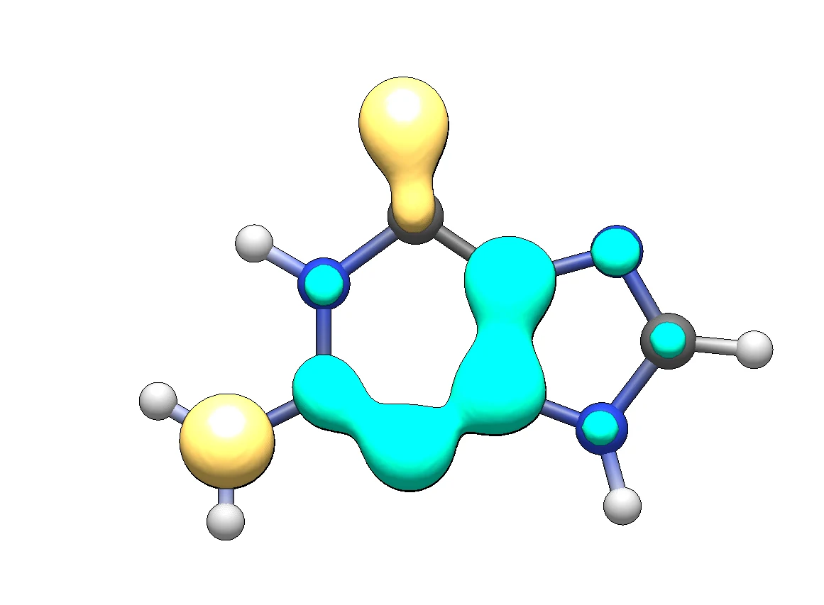
Figure 2. BDFπ of isolated guanine, isosurface 0.015. Turquoise: BDFπ > 0 (delocalized bonding dominates); yellow: BDFπ < 0 (localized electrons).
A delocalized domain runs from C2 through N3 and C4 to C5, while the carbonyl group and the lone pair of the amino nitrogen are localized. About 53 % of the π-electrons are delocalized (Total EDDB_G 7.423 of 14.000 e).
Part 2 — BDFπ of the quartet
In the NOBD table the 28 π-NOBDs arrive as seven near-degenerate quartets, one member per guanine (1.609, 1.532, 1.205, 1.103, 1.007, 0.777, 0.737), followed by a sharp gap to NOBD 29 at 0.176:
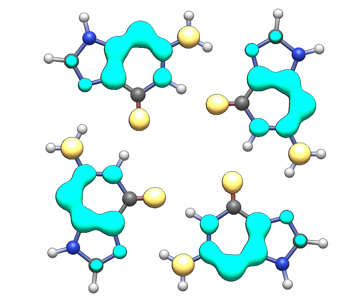
Figure 3. BDFπ of the guanine quartet, isosurface 0.015.
At first sight each guanine keeps the pattern of the isolated molecule. The difference shows in the EDDB_G numbers: the delocalized fraction rises from 53.0 % in guanine to 56.9 % in the quartet (31.882 / 4 − 7.423 = +0.548 e per guanine). The changes are too subtle to read by comparing Figures 2 and 3 by eye. That is what the differential map is for.
Part 3 — The differential ΔBDFπ map
We subtract the reference from the quartet, point by point on the reference grid. Positive regions gained delocalization on forming the quartet; negative regions became more localized.
- --export-cube dbdf — write the differential map, G4.DBDF_G.cube.
- --cube-ref G.BDF_G.cube — the reference; its grid is cloned and its values subtracted.
- --cube-align 1:16 — superimpose the quartet onto the reference frame by a rigid fit of atoms 1–16, the first guanine of G4.xyz. The alignment atoms must have the same index and element in both files.
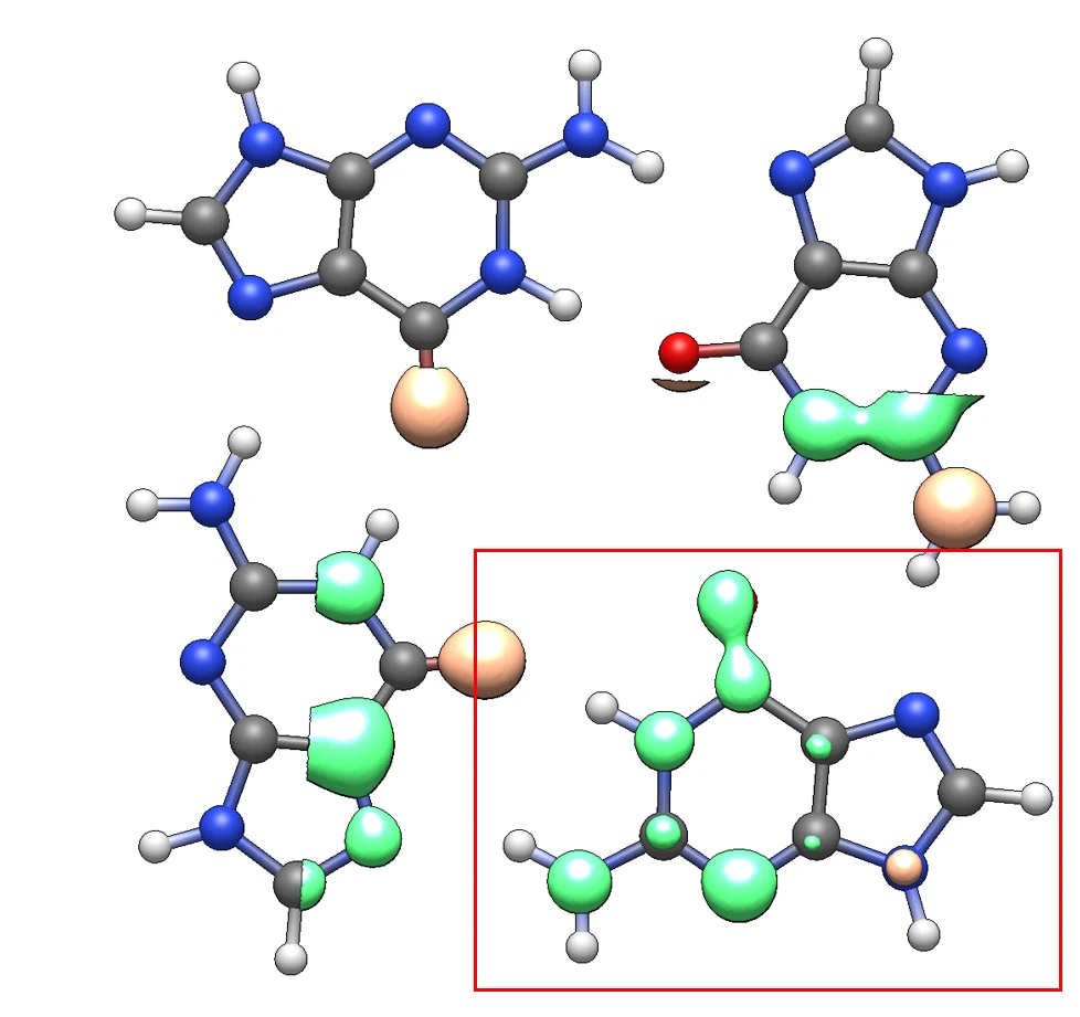
Figure 4. The raw ΔBDFπ map, isosurface ±0.010. Green: ΔBDFπ > 0; orange: ΔBDFπ < 0. Only the guanine in the red box — the one aligned onto the reference — carries a meaningful difference.
The reference holds a single isolated guanine, superimposed on the first guanine of the quartet, so nothing is subtracted at the positions of the other three: the partially visible lobes there are their raw BDFπ in the quartet. And because the map exists only inside the grid box of the monomer, those raw lobes are cut off at the box edge. The only part of the map to read is inside the red box.
Part 4 — The full map from symmetry
All four guanines are equivalent, so the map of the aligned guanine represents the other three. Rotating it by 90°, 180° and 270° about the four-fold axis gives the full ΔBDFπ map:
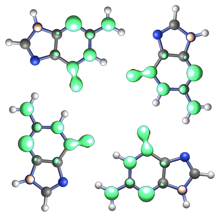
Figure 5. ΔBDFπ for the full quartet, assembled from the aligned guanine by four-fold rotation. Isosurface ±0.010.
- Delocalization increases over the pyrimidine half of each guanine and along the C=O bond.
- The imidazole ring is less affected.
- The only negative lobe is a small one on N9, which becomes slightly more localized.
Part 5 — π-Cloud polarization
The NPA tables of G.log and G4.log give the change of the π-population of every atom, ΔED_G = ED_G(G4) − ED_G(G):
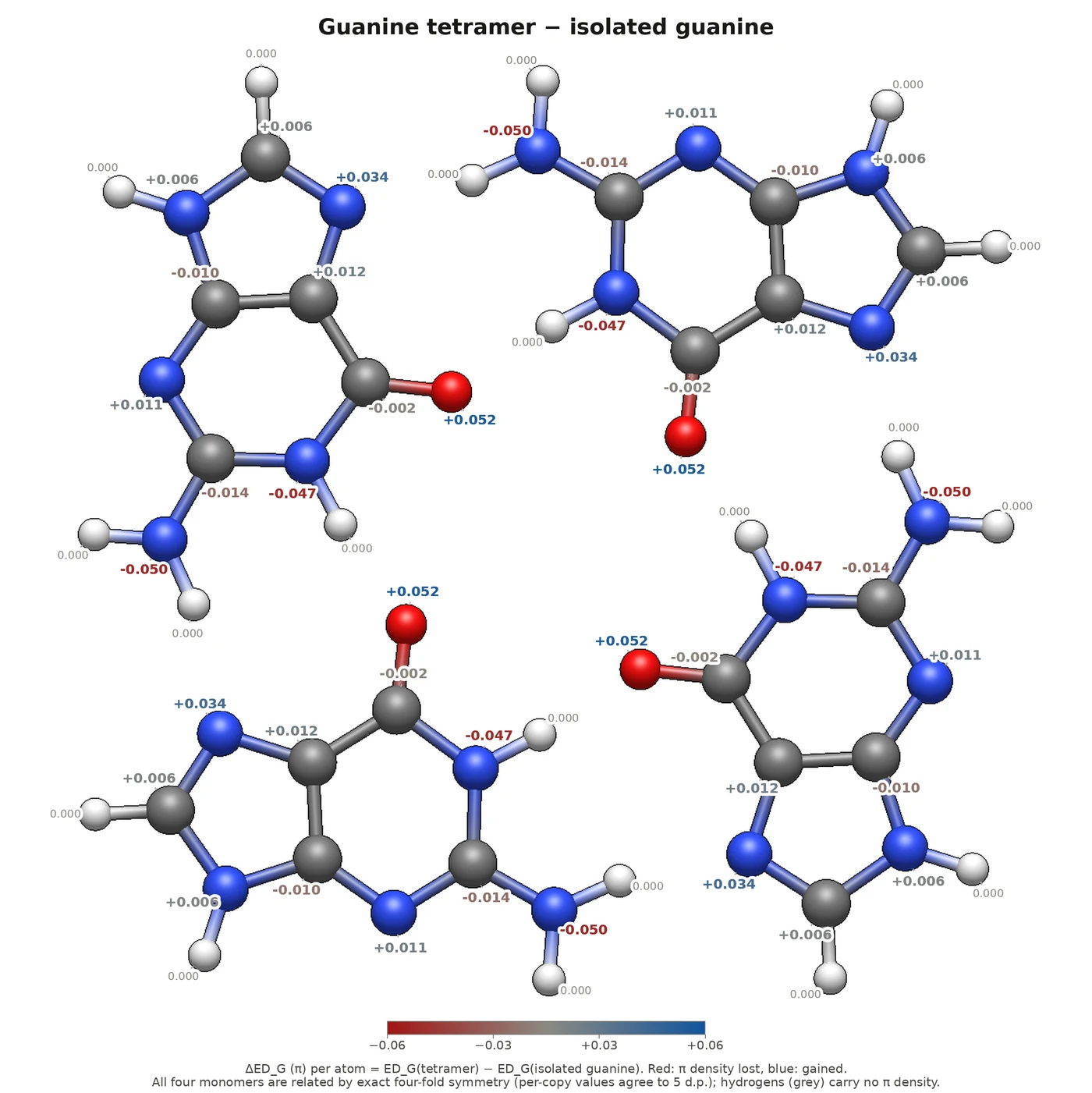
Figure 6. Change in π-electron population on forming the quartet, ΔED_G. Red: π-density lost; blue: gained.
The four largest changes are on the four atoms that take part in hydrogen bonding. The donor nitrogens lose π-density (N1 −0.047 e, N2 −0.050 e) and the acceptors gain it (O6 +0.052 e, N7 +0.034 e): π-density moves from the Watson–Crick edge of each guanine to its Hoogsteen edge. Each guanine still holds exactly 14.000 π-electrons, so the density is redistributed within each guanine, not transferred between them.
Table 1. Changes per guanine on forming the quartet, from the NPA tables of G.log and G4.log. Shaded rows are the hydrogen-bonding atoms; the atom index in the .xyz files is given in brackets. Atom numbering in Figure 7.
| atom | role in the quartet | ΔED_G | ΔEDDB_G | ΔBDF lobe |
|---|---|---|---|---|
| N1 (3) | H-bond donor (N1–H···O6) | −0.047 | +0.054 | green |
| N2, amine (1) | H-bond donor (N2–H···N7) | −0.050 | +0.068 | green |
| O6 (2) | H-bond acceptor | +0.052 | +0.111 | green |
| N7 (11) | H-bond acceptor | +0.034 | +0.025 | — |
| C2 (4) | −0.014 | +0.037 | green | |
| N3 (5) | +0.011 | +0.137 | green | |
| C4 (6) | −0.010 | +0.019 | — | |
| C5 (7) | +0.012 | +0.035 | — | |
| C6 (8) | −0.002 | +0.078 | green | |
| C8 (10) | +0.006 | +0.016 | — | |
| N9 (9) | +0.006 | −0.031 | orange | |
| per guanine | 0.000 | +0.548 |
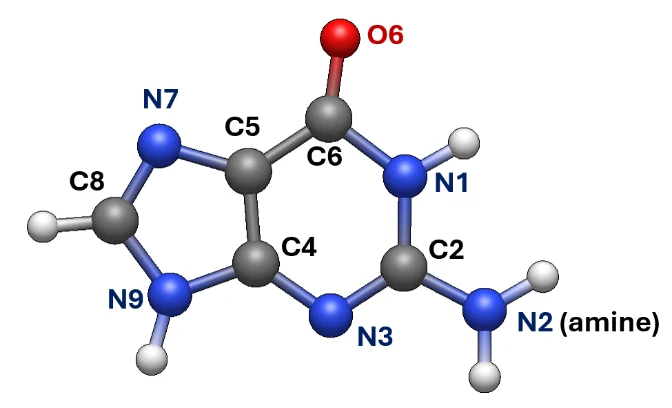
Figure 7. Atom numbering of guanine.
Part 6 — Resonance interpretation
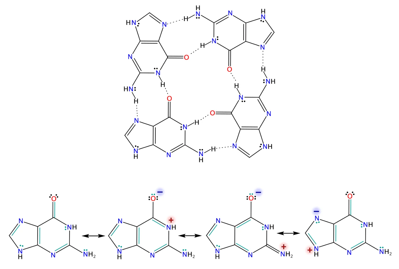
Figure 8. Top: the hydrogen-bond pattern of the quartet. Bottom: resonance structures that explain the changes in delocalization and in the atomic π-populations.
The second and third structures carry the lone pairs of N1 and of the amino nitrogen N2 onto O6. They account for the donors losing π-density, O6 gaining it, and the C6=O6 bond becoming more delocalized — the green dumbbell on the carbonyl in Figure 5. Hydrogen bonding at the donor edge and at O6 favours exactly these charge-separated forms.
The fourth structure places the negative charge on N7, through a charge transfer from N9. Why then does the N9 lone pair become slightly more localized (the orange lobe in Figure 5)? Because this resonance competes with the cyclic delocalization in the only aromatic ring of guanine, the imidazole ring. Its CDI (see Tutorial 1 and Tutorial 2) is lower in the quartet than in the isolated guanine. Run the pathway analysis for the five-membered ring (atoms 6-7-11-10-9 of the first guanine) in both files:
The CDI drops from 0.410 (G) to 0.377 (G4), while the six-membered ring (-p 3-4-5-6-7-8-3) goes the other way, from 0.157 to 0.207. The cyclic delocalization of the imidazole ring also runs through resonance structures with charge transfer from the N9 lone pair, so the overall delocalization of that lone pair can decrease even though one single contributor grows.
How much this π-reorganization contributes to the strength of the hydrogen bonds is a separate question. Energy decomposition analyses of G-quartets attribute their cooperativity mainly to σ donor–acceptor interactions, with only a small π contribution [C. Fonseca Guerra, H. Zijlstra, G. Paragi, F. M. Bickelhaupt, Chem. Eur. J. 2011, 17, 12612]. The differential map shows clearly that a π-response exists; it does not by itself measure its energetic weight.
What to try next
- Export deddb and dedlb instead of dbdf and see which component carries the change.
- Check the symmetry argument of Part 4: reorder G4.xyz so that the second guanine (atoms 17–32) comes first, align on atoms 1–16 again, and compare the two differential cubes. (--cube-align 17:32 itself would fail: the reference has only 16 atoms, and the alignment atoms need the same index in both files.)
Tutorial prepared by Paweł Wieczorkiewicz.